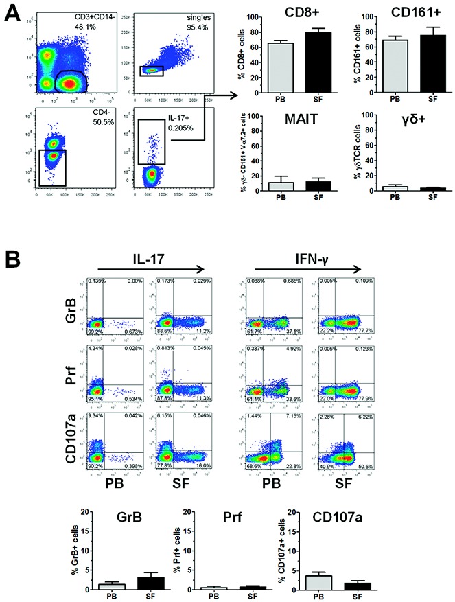
Figure 2.

CD3+CD4− T cells in patients with psoriatic arthritis (PsA) are predominantly CD8+ T cells lacking markers associated with cytolytic activity. Cryopreserved mononuclear cells from paired peripheral blood (PB) and synovial fluid (SF) samples from patients with PsA were thawed and stimulated with phorbol myristate acetate and ionomycin in the presence of GolgiStop for 3 hours, and then stained for the expression of CD3, CD4, CD8, CD161, γ/δ T cell receptor (TCR), Vα7.2 TCR, granzyme B (GrB), perforin (Prf), and CD107a, along with interleukin-17 (IL-17). A, Left, Gating strategy to identify IL-17+CD3+CD4− T cells by flow cytometry. Representative results are shown. A, Right, Percentage of cells within the IL-17+CD3+CD4− T cell population that were either CD8+, CD161+, γ/δ−CD161+Vα7.2+ (mucosal-associated invariant T [MAIT] cells), or γ/δ+. B, Top, Coexpression of IL-17 or interferon-γ (IFNγ) and granzyme B, perforin, or CD107a in CD3+CD8+ T cells from PsA PB and SF, as determined by flow cytometry. Representative dot plots are shown. B, Bottom, Percentage of cells within the IL-17+ CD3+CD8+ T cell population that expressed granzyme B, perforin, or CD107a in PsA PB and SF. Results are the mean ± SEM of 4 samples. Color figure can be viewed in the online issue, which is available at http://onlinelibrary.wiley.com/doi/10.1002/art.38376/abstract.
