Figure 5.

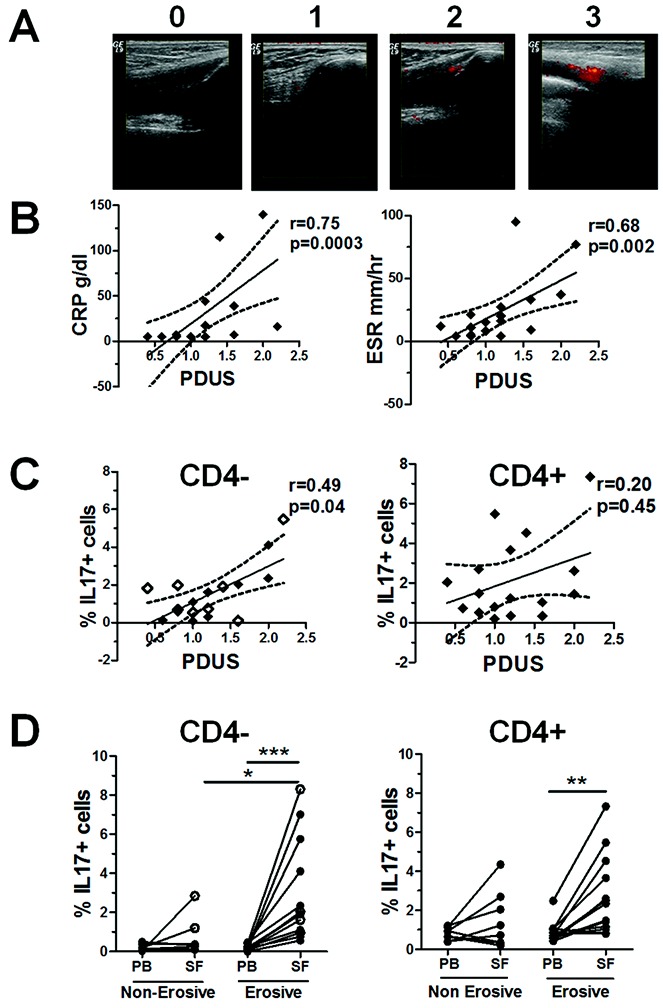
Correlation between the frequency of interleukin-17 (IL-17)–expressing CD4− T cells and the power Doppler ultrasound (PDUS) score for the presence of local synovitis in patients with psoriatic arthritis (PsA), and enrichment of this T cell subset in the joints of patients with erosive disease. A, Representative PDUS images illustrating the semiquantitative grading system for active synovitis in the knee joints of patients with PsA, where 0 = no signal, 1 = 1–2 pixels, 2 = <50% signal, and 3 = ≥50% signal. B, Correlations between measures of disease activity (the C-reactive protein level or erythrocyte sedimentation rate) and the mean PDUS score of the aspirated knee joints (n = 17). C, Correlations between the percentage of IL-17+ cells within CD3+CD4− T cells (including CD3+CD8+ T cells [open diamonds]; n = 8) or CD3+CD4+ T cells from PsA synovial fluid (SF) and the mean PDUS score in the same knee joint. In B and C, regression coefficients (solid lines) with 95% confidence intervals (broken lines) are shown for each plot. D, Percentage of IL-17+ cells within the CD3+CD4− T cell (including CD3+CD8+ cells [open circles]) and CD3+CD4+ T cell populations in paired samples of peripheral blood (PB) and SF from patients with erosive PsA (n = 13) compared to patients with nonerosive PsA (n = 8). Each symbol joined by a line represents a paired sample from a different patient. Data were analyzed using one-way analysis of variance for parametric or nonparametric data, where appropriate. ∗ = P < 0.05; ∗∗ = P < 0.01; ∗∗∗ = P < 0.001.
