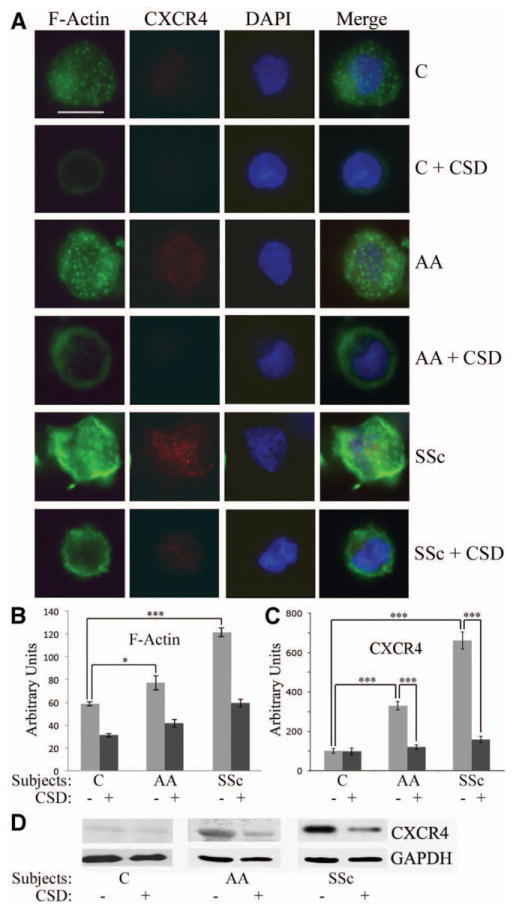
Figure 2.

The CSD peptide reverses the enhanced expression of F-actin and CXCR4 observed in monocytes from African Americans and patients with SSc. Monocytes were isolated from the blood of healthy Caucasians and African Americans, and from patients with SSc. One portion was attached to coverslips, treated with CSD or control peptide, stained for F-actin and CXCR4, and counterstained with DAPI. Another portion was treated with CSD or control peptide and extracted for Western blotting. A, Representative images showing F-actin and CXCR4 expression in cells obtained from healthy Caucasians and African Americans and from patients with SSc (n = 4 donors in each group), with and without CSD peptide treatment. Staining for F-actin and CXCR4 was enhanced in monocytes from healthy African Americans and from patients with SSc, and treatment with CSD decreased this staining. Bar = 5 μm. B and C, Densitometric quantification of staining for F-actin (B) and CXCR4 (C). Values are the mean ± SEM (n = 10 cells in each group). * = P < 0.05; *** = P < 0.001. D, Representative Western blot showing enhanced CXCR4 expression in monocytes from African Americans and patients with SSc that was reversed by CSD treatment. The level of CXCR4 in Caucasian subjects treated with control peptide was set at 100 arbitrary units. See Figure 1 for definitions.
