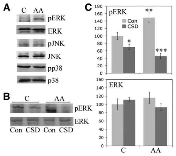
Figure 3.

Enhanced ERK signaling in monocytes from African Americans. Monocytes were isolated from the blood of healthy Caucasian and African American donors and cultured on 6-well plates. The expression and activation of the indicated signaling molecules were evaluated by Western blotting (50 μg total protein per lane). A, Representative set of Western blots showing hyperactivation of ERK, but not JNK or p38, in monocytes from African Americans compared with those from Caucasians. Similar results were obtained in 3 experiments performed with cells from different subjects. B, Representative Western blot showing that CSD peptide reverses the enhanced activation of ERK in monocytes from African Americans incubated with control (Con) peptide. C, Quantification of the results shown in B, as determined by densitometry. The levels of pERK and ERK in Caucasian subjects treated with control peptide were set at 100 arbitrary units. Values are the mean ± SEM of 3 independent experiments using cells from different subjects. * = P < 0.05 versus monocytes from Caucasians treated with control peptide; ** = P < 0.01 versus monocytes from Caucasians treated with control peptide; *** = P < 0.001 versus monocytes from African Americans treated with control peptide. See Figure 1 for other definitions.
