Figure 6.

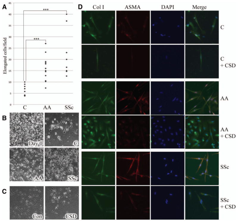
Enhanced monocyte-to-fibrocyte differentiation in healthy Caucasian and African American donors and in patients with SSc. A, Fibrocyte differentiation as quantified by the number of peripheral blood mononuclear cells (PBMCs) that acquire a spindle-shaped morphology in culture. Each data point represents a single subject. *** = P < 0.001. B, Representative image showing a typical PBMC starting population (day 0) (top left) and representative fields showing fibrocyte differentiation in PBMCs from healthy Caucasians (top right) and African Americans (bottom left) and patients with SSc (bottom right) after 15 days of culture. C, Inhibition of fibrocyte differentiation by CSD peptide. PBMCs from Caucasian donors were treated with control (Con) peptide or CSD peptide during 15 days of culture. Similar inhibition of fibrocyte differentiation by CSD was observed in 5 experiments performed with PBMCs from different healthy Caucasian and African American donors and patients with SSc. D, Fibrocyte phenotypes. Fibrocytes from healthy Caucasian and African American donors and patients with SSc were stained with antibodies against type I collagen (Col I) and α-smooth muscle actin (α-SMA) and counterstained with DAPI. Note that α-SMA staining was more prominent in fibrocytes from African Americans and patients with SSc compared with that in fibrocytes from Caucasians and was inhibited by CSD treatment. Similar results were obtained in 3 experiments performed with PBMCs from different Caucasian and African American donors and patients with SSc. Original magnification × 200. See Figure 1 for other definitions.
