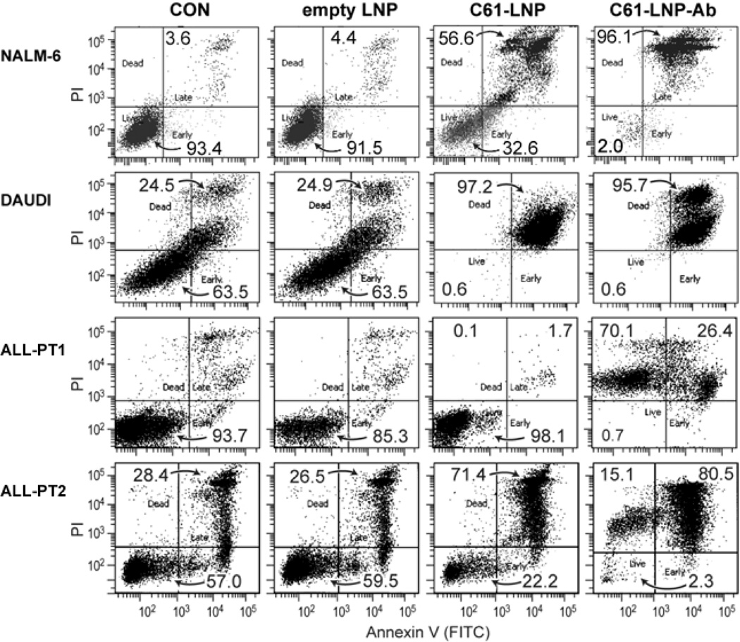
Figure 6. In Vitro Anti-leukemic Potency of Unmodified and Antibody-Decorated C61-LNP Against Leukemia Cell Lines and Primary B-precursor ALL Cells.
Representative two-color fluorescence density plots of leukemia cell lines (NALM-6 and DAUDI) and primary leukemia cells from 2 pediatric B-precursor ALL patients (ALL-PT1 and ALL-PT2). The PI fluorescence intensity is depicted along the Y-axis and the Annexin V (FITC) fluorescence is depicted along the X-axis. Cells were treated for 48 h at 37°C with unmodified (C61-LNP, 30 µg/mL) or antibody-decorated C61-LNP (C61-LNP-Ab, 30 µg/mL). Controls included sham-treated cells (CON) and cells treated with empty LNP (C61-free empty LNP with the same lipid content as C61-LNP used at 30 µg/mL C61-based concentration). Cells were analyzed for apoptosis using the standard quantitative flow cytometric apoptosis assay using the Annexin V-FITC Apoptosis Detection Kit from Sigma as discussed in the Methods section. The labeled cells were analyzed on a LSR II flow cytometer. C61-LNP-Ab caused apoptosis in the vast majority of treated B-precursor ALL cells. The anti-leukemic potency of C61-LNP is evidenced by the significantly lower percentages of Annexin V-FITC−PI− live cells located in the left lower quadrant of the corresponding two-color fluorescence density plots as well as a substantially higher percentage of Annexin V-FITC+PI+ apoptotic cells located in the right upper quadrant. The cumulative results are depicted in Figure 7.

