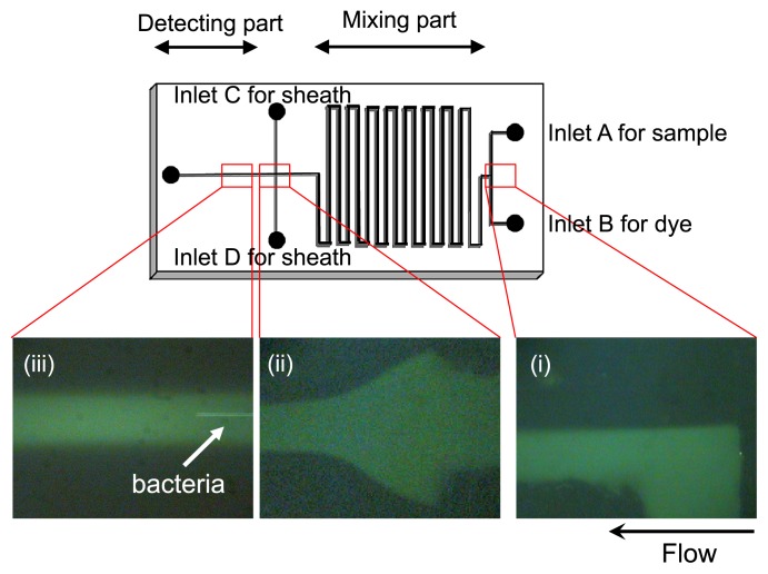
Fig. 5.
Details of the microfluidic device for on-chip staining and counting of bacterial cells (size: 5 cm × 2.5 cm). (i) Samples and fluorescent dye solution flow separately and are then mixed through the “mixing part” of the microchannel. (ii) Alignment of sample flow by sheath fluid. (iii) Flow of bacterial cells in the “detecting part” of the microchannel.

