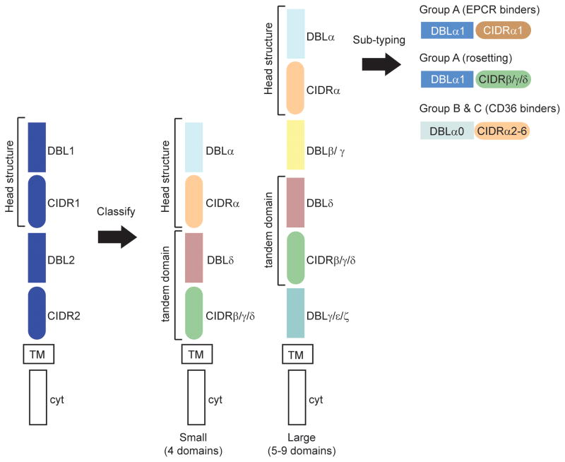
Fig 1.
Adhesion domain classification of PfEMP1 proteins. The blue PfEMP1 shows a typical arrangement of PfEMP1 domains. The first arrow indicates how adhesion domain classification reveals higher domain organization in PfEMP1. Specific DBL and CIDR domain types form preferential tandem domain arrangements (DBLα-CIDRα and DBLδ-CIDRβ/γ/δ). The same tandem arrangements are present in small (4-domain) and large (5 or more domain) PfEMP1, but large proteins contain unique DBL subtypes (β or γ) that are not present in small proteins. The second arrow indicates that further sub-classification of adhesion domains (e.g. CIDRα1) distinguishes three different PfEMP1 head structure types and functional differences between group A, B, and C proteins. TM is transmembrane, cyt is cytoplasmic tail.

