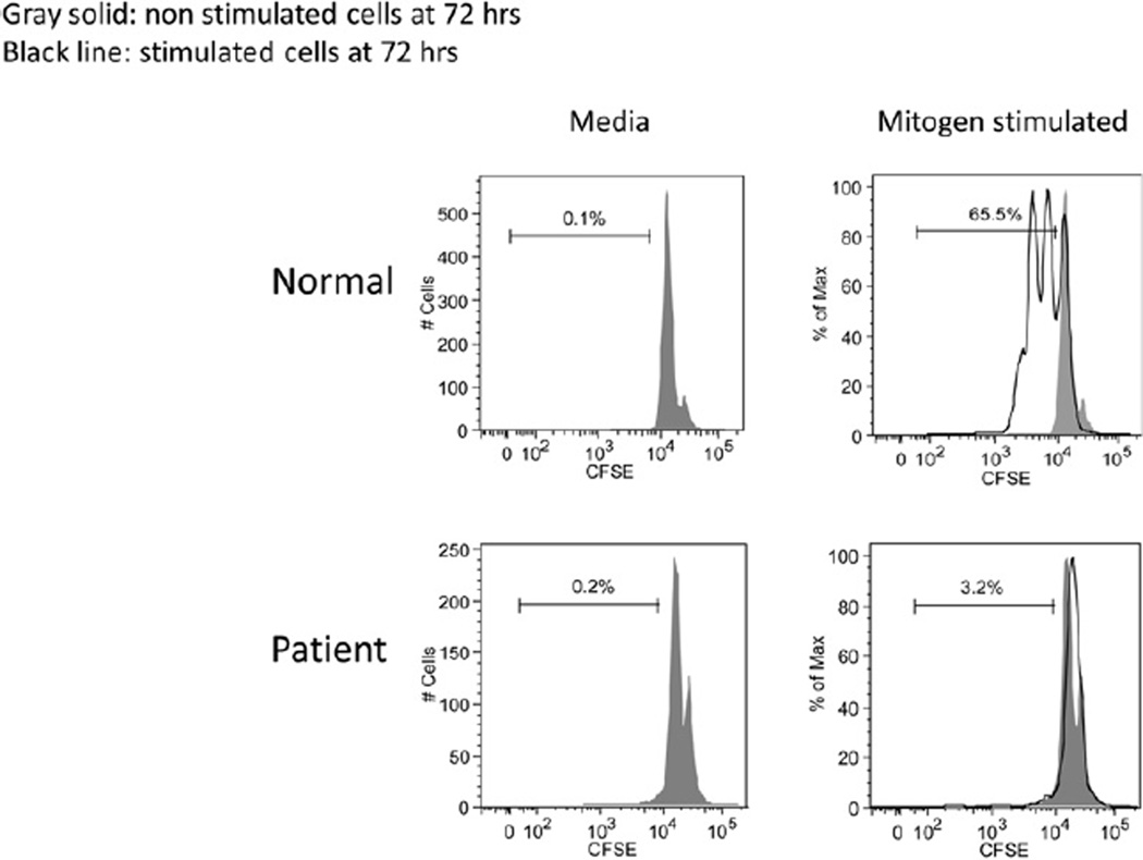
FIG E1.
Flow cytometry–based lymphocyte proliferation assay using a cell-tracking dye. Cells were incubated with CFSE, a fluorescent dye that binds covalently to cytosolic proteins, and then stimulated with mitogens. CFSE dilution could be detected on the control cells after cell division/proliferation but not on the patient’s cells.

