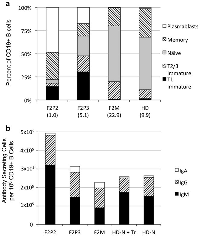
Fig. 2.
B cell Phenotype: a Peripheral blood B cell subtypes in affected patients (F2P2 and F2P3) and unaffected mother (F2M) versus healthy donor (HD) controls as measured using flow cytometry of isolated peripheral blood mononuclear cells (PBMCs). Numbers in parenthesis represent the percentage of CD19+ B cells out of total PBMCs. B cell subsets were defined using cell surface markers as specified by Moir, et. al. (2013) [10]. b Number of antibody-secreting cells measured by ELISPOT (spots per 106 CD19+ B cells). PBMCs from Patients (F2P2 and F2P3) and unaffected mother (F2M) were stimulated in vitro with S. aureus Cowan strain I (SAC), CpG, IL-21 and pokeweed mitogen (PWM) for 4 days. CD19-depleted PBMCs from a healthy donor (HD) reconstituted with naïve (N) or naïve and transitional (N + Tr) B cells (CD27/IgG-negative fraction of CD10-depleted or CD10-containing B cells respectively of a donor whose CD10+ transitional B cells represented 20 % of total B cells) were included. Results for healthy donor cells were representative of three independent replicates

