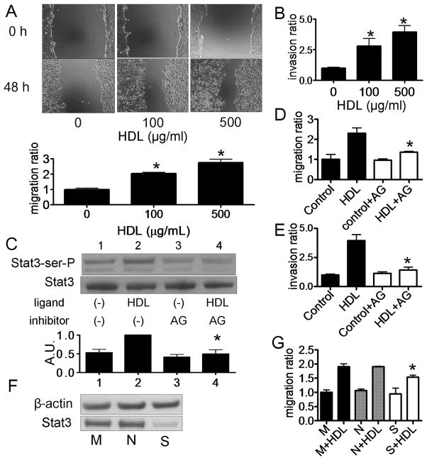
Fig. 2.
Migration and invasion of DU145 cells by HDL via Stat3 activation. A, D: A monolayer of DU145 cells were scored and then cultured in medium for 0 or 48 h with or without HDL. Cell migration into the wound was examined by phase-contrast microscopy. Cells were preincubated in the absence (−) or presence of AG490 (50 μM) for 16 h before HDL treatment (Panel D). Values are expressed as the mean +SD (n=3). *P<0.01 vs 0 μg/mL. AG; AG490. B, E: After culturing DU145 cells with HDL, the numbers of cells that invaded through the matrigel were counted at the microscope. Cells were preincubated in the absence (Control) or presence of AG490 (50 μM) for 16 h before HDL treatment (Panel E). Values are expressed as the mean +SD (n=3). *P<0.05 vs 0 μg/mL (Panel B), *P<0.05 vs HDL (Panel E). C: DU145 cells were preincubated in the absence (−) or presence (+) of AG490 (50 μg/mL) for 16 h, and then incubated with or without HDL (500 μg/ml) for 15 min. Whole cell lysate was analyzed by western blotting. The graph shows the ratio of pStat3 to Stat3 in each sample relative to 1.0 for lane 2 (n=3, mean +SD). *P<0.05 vs lane 2. AU; arbitrary unit F: Effect of siRNA for Stat3 expression in DU145 cells was evaluated. Mock transfected cells (M) or cells transfected with Stat3 siRNA (S) or negative control siRNA (N), cells were incubated for 48 h before harvest for western blotting. G: After transfection of siRNA, DU145 cells were incubated with the medium containing 10% FBS for 24 h, and medium was switched to serum-free medium. After 24 h, cells were wounded and then cultured for 0 or 48 h with or without HDL (500 μg/mL). Cell migration into the wound was examined by phase-contrast microscopy. Values are expressed as the mean +SD. (n=3). *P<0.05 vs N+HDL. M; mock, N; negative and S; Stat3.

