Fig. 5.
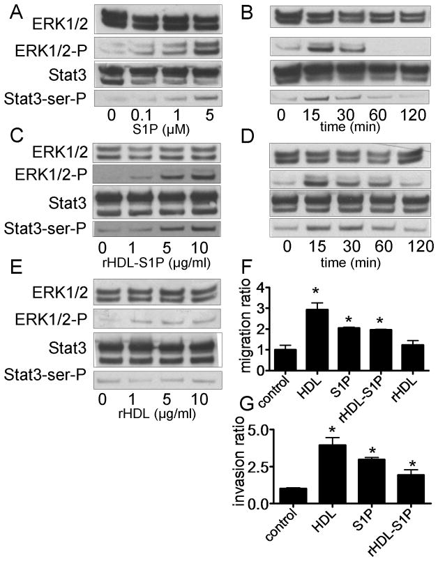
Stat3 activation by S1P, rHDL-S1P and rHDL. A-E: DU145 cells cultured in medium without FBS for 24 h were analyzed by western blotting after 15 min (Panel A, C and E) for the indicated concentration of S1P (Panel A), rHDL-S1P (Panel C) or rHDL (Panel E), or after treatment for the indicated times with S1P (Panel B; S1P 5 μM) or rHDL-S1P (Panel D; 10 μg/ml). F: DU145 cells were wounded and cultured in the medium for 0 or 48 h with or without S1P (5 μM), rHDL-S1P (10 μg/ml), or rHDL (10 μg/ml). Cell migration into the wound was examined by phase-contrast microscopy. Values are expressed as the mean +SD (n=3). *P<0.01 vs control. G: After culturing DU145 cells with S1P (5 μM) or rHDL-S1P (10 μg/ml), the numbers of cells that went through the matrigel were counted at the microscope. Values are expressed as the mean +SD (n=3). *P<0.05 vs control.

