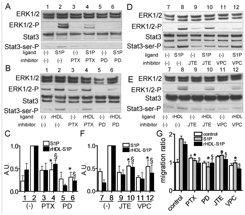
Fig. 6.
Stat3 activation by S1P and rHDL-S1P via S1P2 and S1P3 in DU145 cells. A-F: DU145 cells were preincubated in the absence (−) or presence of PTX (100 ng/mL) for 24 h, PD98059 (2.5 μM) for 1 h, JTE013 (5 μM) for 1 h or VPC23019 (5 μM) for 1 h. Cells were incubated with or without S1P (5 μM) (Panel A and B) or rHDL-S1P (10 μg/ml) (Panel D and E) for 15 min, and whole cell lysate was analyzed by western blotting. The graph shows the ratio of Stat3-ser-P to Stat3 in each sample relative to 1.0 for lane 2 (n=3, mean +SD). The results of Panel A and B, and Panel D and E were shown in Panel C and D, respectively. *P<0.05 vs lane 2 of S1P, §P<0.05 vs lane 2 of rHDL-S1P. G: Cells were wounded and then cultured for 0 or 48 h with or without S1P (5 μM) or rHDL-S1P (10 μg/ml). Cell migration into the wound was examined by phase-contrast microscopy. Values are expressed as the mean +SD. *P<0.05 vs control of S1P, §P<0.05 vs. control of rHDL-S1P.

