
Fig. 2.
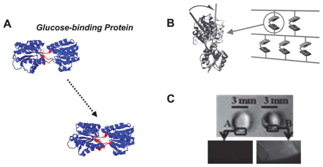
(A) Glucose binding protein (GBP) crystal structures before and after binding to glucose (PDB: 2fw0 → 2fvy). A central region is shown in red as a point of reference to demonstrate the hinge motion. (B) Schematic representing design of poly(acrylamide) hydrogels that include GBP. (C) Top view of a cylindrical hydrogel before and after glucose binding, demonstrating GBP’s dynamic function (B and C from Ehrick et al.41, copyright Wiley-VCH Verlag GmbH & Co. KGaA. Reproduced with permission).