
FIG. 3.
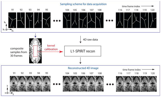
A schematic of the proposed data sampling and image reconstruction. Golden-angle radial spokes sampling in (ky, kz) space is shown in the top block. In the sampling, each frame has 100 (ky,kz) samples along 3, 4, or 5 radial spokes, and each frame shows discontinuity in some spokes which is connected when considering samples from previous or next frames. The composite samples contain fully sampled low-resolution data that are used for the kernel calibration in L1-SPIRiT. L1-SPIRiT reconstruction results in time-resolved 3D volume of the pharyngeal airway as indicated in the bottom block. [Color figure can be viewed in the online issue, which is available at wileyonlinelibrary.com.]