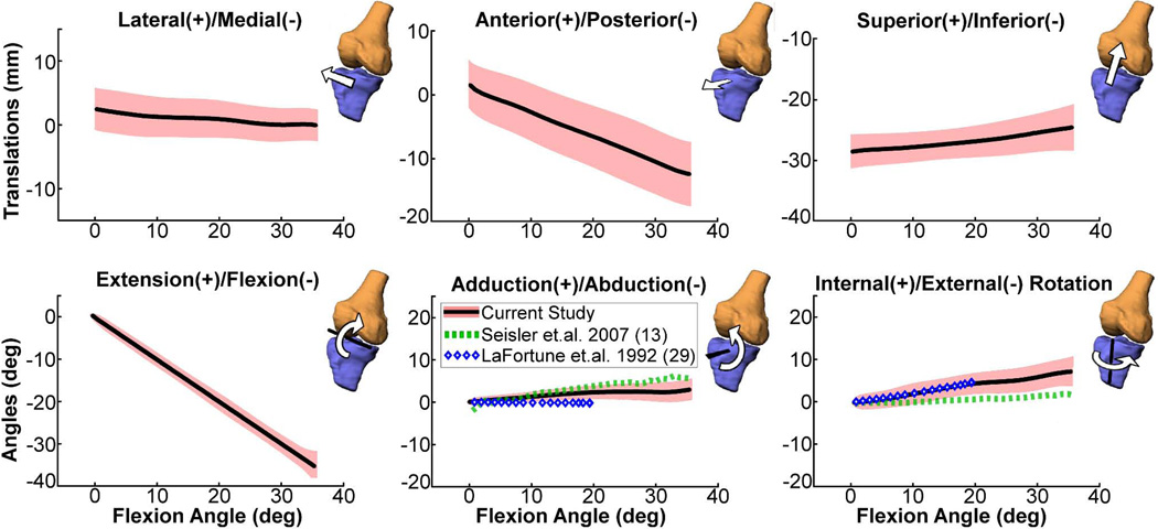
Figure 6.
Ensemble average tibiofemoral kinematics (shaded curves represent mean±1 s.d.) for the dominant knee of ten asymptomatic subjects over a flexion-extension motion cycle as measured using SPGR-VIPR. For comparison, the average adduction and rotation angle data measured by cine-PC MRI during unloaded knee flexion-extension (13) and via intra-cortial traction pins during normal gait (29) are shown. Our data shows greater internal tibia rotation with knee flexion compared to cine PC, which may be related to greater quadriceps activation induced by our loading device (35).

