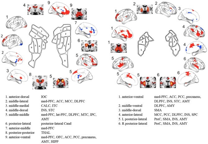
Figure 7. Summary of striatal subdivisions and their functional connectivity maps.
The schematic illustrates parcellations based on the optimal K (9 and 6 for the caudate and the putamen respectively). Regions showing significant functional connectivity to each striatal cluster are depicted on the surface and the section of the brain, and are also listed. IOC, inferior occipital cortex; med-PFC, medial prefrontal cortex; ACC, anterior cingulate cortex; MCC, middle cingulate cortex; DLPFC, dorsolateral prefrontal cortex; CALC, calcarine cortex; ITC, inferior temporal cortex; INS, insula; STC, superior temporal cortex; lat-PFC, lateral prefrontal cortex; MTC, middle temporal cortex; IPC, inferior parietal cortex; AMY, amygdala; Caud, caudate; THAL, thalamus; OFC, orbitofrontal cortex; PCC, posterior cingulate cortex; HIPP, hippocampus; SMA, supplementary motor area; SPC, superior parietal cortex; PreC, precentral cortex.

