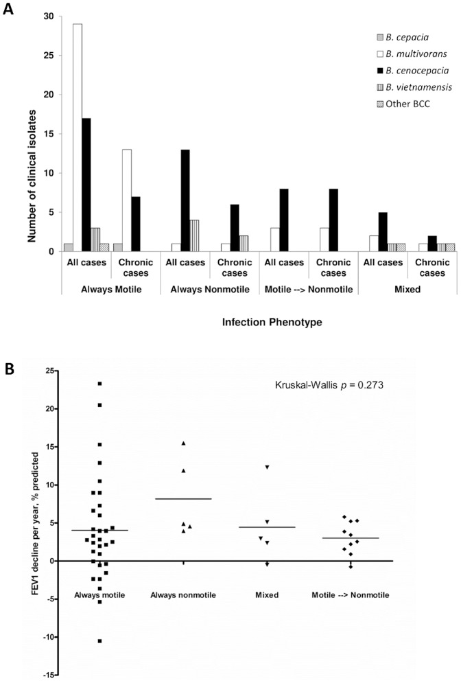
Figure 3. Assessment of clinical significance of swimming motility in BCC bacteria.
A) CF lung infections categorized by species and motility phenotype. Data are displayed for all cases (n = 89) and for cases where chronic infection (n = 48) was clearly established (defined as the presence in our collection of at least 2 or more isolates of the same strain type that were collected at least a year apart). Grey bars = B. cepacia, white bars = B. multivorans, black bars = B. cenocepacia; vertical striped bars = B. vietnamiensis and diagonal striped bars = other BCC species. Infections were categorized as: isolates were always motile; always nonmotile; motile to nonmotile phenotype switches detected during infection (as defined by an initial isolation of at least 2 separate motile isolates on separate months followed by a nonmotile isolate at any point thereafter) or mixed (there was no clear established initial swimming motility phenotype as the first two isolates stored for patients on separate months demonstrated both motile and nonmotile phenotypes). One case was also classified as mixed (FEV1% predicted decline −0.5%) as most isolates were nonmotile, however several marginally motile (diameter 11 and 12 mm) isolates were detected during the course of the patients' infection. B) Assessment of the impact of motility phenotype on FEV1 decline following initial infection with BCC. FEV1 data were collected from the patient charts as described previously. Criteria for inclusion were established previously [22]. Data were analyzed in GraphPad Prism v.4 and the non-parametric Kruskal-Wallis test calculated to assess difference.

