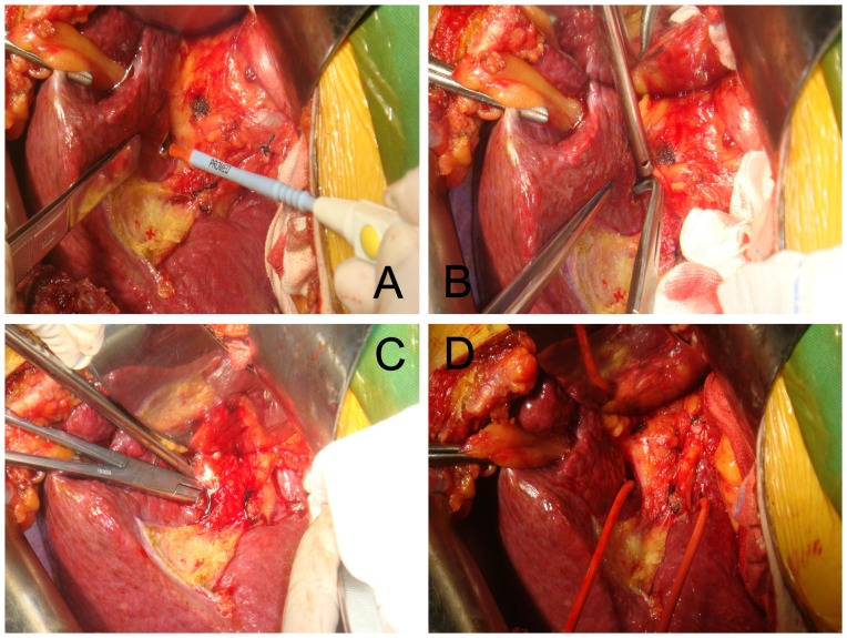
Figure 1. Simple hemi-occlusion.
A: On the visceral envelope overlying the confluence, a small hole was made using a sharp blade; B: A right-angle forceps was inserted to gently mobilize the liver parenchyma outside Glisson’s sheath; C: The right-angle forceps should mobilize in the liver parenchyma towards the caudate lobe; D: A catheter was introduced.

