Figure 2.

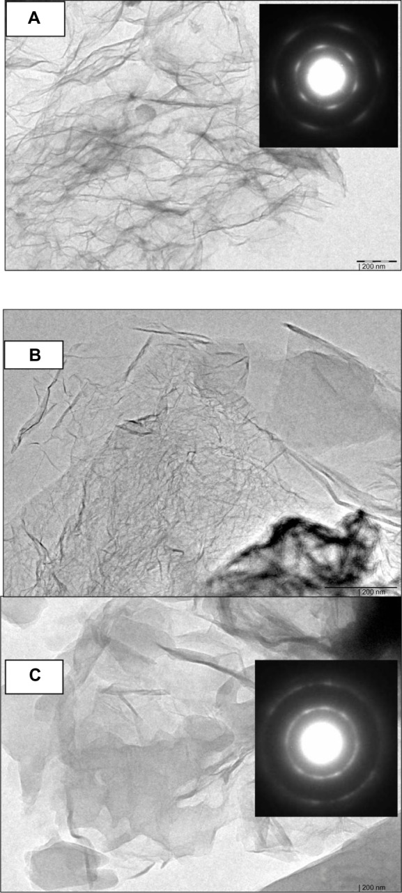
TEM images of GS (A), f-Gr (B), and f-Gr-AmB (C).
Notes: The TEM image of GS shows a wrinkled paper-like structure (A), which appears to be very thin. The inset is the selected area electron diffraction pattern (SAED) of GS, showing clear diffraction spots; the diffraction spots were indexed to a hexagonal graphite crystal structure. In contrast, the TEM image of f-Gr (B) shows a smooth surface, which is due to functionalization by L-cysteine. AmB deposited on f-Gr (C) appears as a cloudy shadow. The SAED pattern of the inset demonstrates the well-textured, crystalline nature of f-Gr-AmB, which supports the conclusion that AmB is attached.
Abbreviations: TEM, transmission electron microscopy; GS, graphene sheets; f-Gr, amine-modified graphene; AmB, amphotericin B; f-Gr-AmB, a novel AmB formulation as a conjugate with f-Gr.
