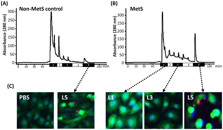
Figure 1. Distribution of LDL subfractions in metabolic syndrome (MetS) and healthy control subjects and the effects of LDL subfractions from MetS subjects on cell death.
Representative chromatographs showing the distribution of LDL subfractions L1–L5 (labeled 1–5) in LDL from a (A) control subject and (B) MetS subject. (C) Effects of L1, L3, and L5 (50 µg/mL each) from MetS subjects and L5 (50 µg/mL) from non-MetS control subjects on bovine aortic endothelial cell (BAEC) death after 24 hours, as determined by staining with Hoechst 33342 (to assess nuclear morphology, blue) and calcein acetoxymethyl ester and propidium iodide (to assess membrane integrity, red). As a negative control, BAECs were incubated with phosphate-buffered saline (PBS) for 24 hours. BAECs that have condensed, fragmented nuclei were considered to be undergoing cell apoptosis.

