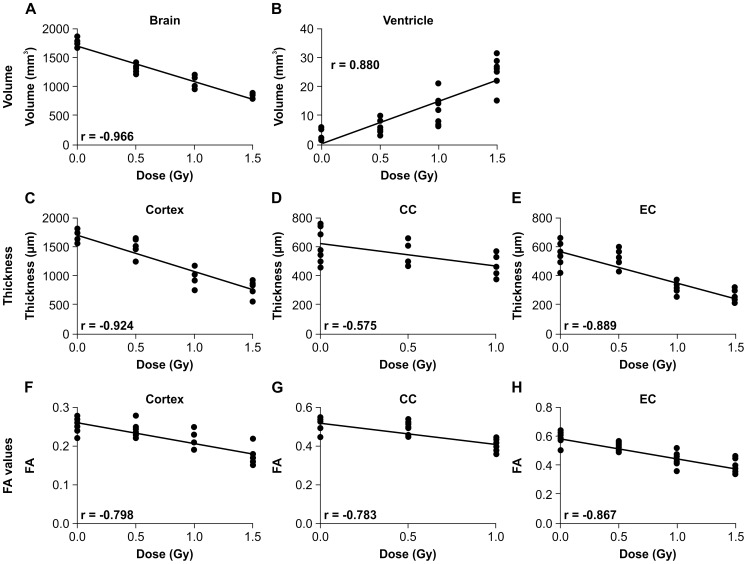
Figure 6. Correlation analysis: correlations of MRI-determined brain volume, ventricle volume, brain substructure thicknesses, and brain substructure FA (fractional anisotropy) values with radiation dose were evaluated.
A, B; Correlation between radiation dose (0.5, 1.0, or 1.5 Gy) and brain volume (A) and ventricle volume (B). C, D, E; Correlation between dose and thickness of cortex (C), thickness of corpus callosum (D), and thickness of external capsule (E). F, G, H; Correlation between dose and FA values of cortex (F), corpus callosum (G), and external capsule (H).

