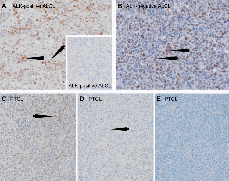
Figure 2.
Single immunoperoxidase labeling demonstrating pFADD expression in ALCL and PTCL (NOS).
Notes: (A) An example of a case of ALK-positive ALCL where the tumor cells express pFADD (arrowed). Note the presence of pFADD in the cytoplasm of mitotic cells (arrowhead). The inset shows an example of a pFADD-negative case of ALK-positive ALCL. (B) Strong nuclear pFADD expression is also present in a case of ALK-negative ALCL (arrowed). Cytoplasmic pFADD is also present in a mitotic cell (arrowhead). Different examples of pFADD in three cases of PTCL are shown in (C–E). While the majority of tumor cells are pFADD-positive (arrowed) in (C), the percentage drops to less than 30% in (D) while the PTCL in (E) lacks detectable pFADD.

