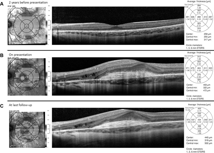
Figure 2.
Spectral domain optical coherence tomography horizontal line scan of the left eye prior to development of vision loss, at presentation, and at last follow-up.
Notes: Intact outer retinal structures centrally with normal foveal contour and peripheral outer retinal and retinal pigment epithelium atrophy (A). Subretinal hyper-reflective material in the central macula consistent with fibrosis with trace subretinal fluid and mild intraretinal fluid overlying an area of retinal pigment epithelium and choroidal atrophy nasally (B). Mild improvement in nasal intraretinal fluid and subretinal fibrosis with resolution of subretinal fluid after four intravitreal bevacizumab injections (C).
Abbreviations: min, minimum; max, maximum; vol, volume; ETDRS, Early Treatment Diabetes Retinopathy Study.

