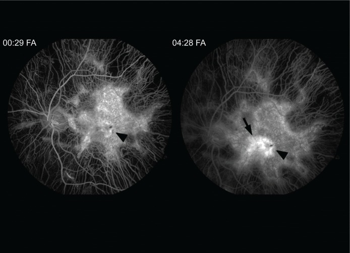
Figure 3.
Fluorescein angiogram (FA) transiting the left eye reveals diffuse atrophy of the choriocapillaris sparing the central macula.
Notes: A central hyperfluorescent lesion in the early images stains (00:29) in the late angiographic images centrally (04:28) (arrowhead). Mild leakage is apparent nasally (arrow).

