Figure 1.
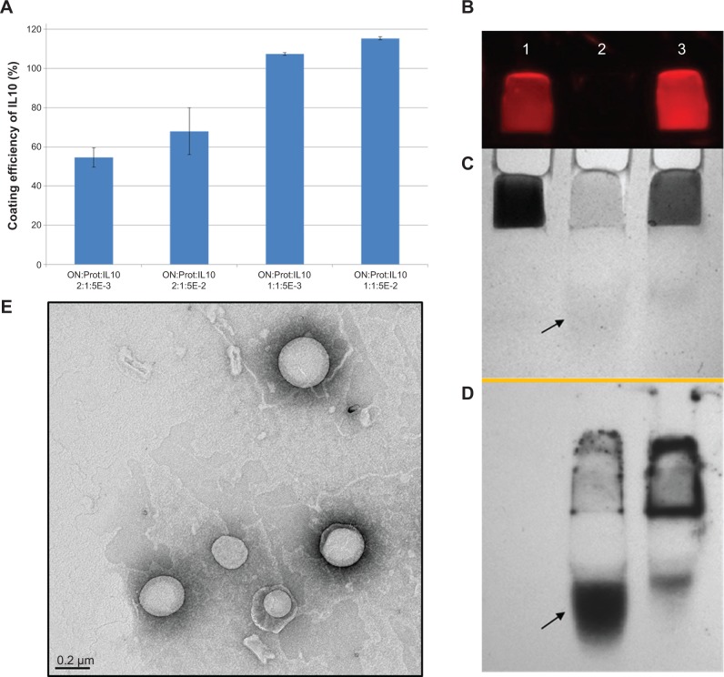
Characterization of IL10 coupled nanoparticles.
Notes: (A) Coating efficiency of mouse IL10. Calculation of coating efficiency as the percentage of deployed IL10. Mean values are shown with error bars representing the standard deviation, n=4. Proticles with a mass ratio of 2:1 (ON:protamine) showed similar IL10 coating efficiencies using different IL10 concentrations. For 1:1 proticles, complete binding of IL10 was recorded, but because the proticles aggregated, IL10 was also incorporated between the aggregates, and thus, not solely presented at the surface. (B–D) IL10-coupled liposomes were applied to a native PAGE gel and then transferred onto a WB membrane. (B) Slots of the gel containing the immobilized probes, visualized by fluorescence imaging when excited at λflu =750 nm. (C) The same gel in (B), but stained with Coomassie blue protein solution, showing that liposomes are stained by the solution. (D) WB membrane of the gel, detected by a primary anti-IL10 Ab and a secondary HRP-conjugated Ab, followed by a substrate reaction and luminescence visualization which shows free and liposomal-bound IL10. Lane 1, Atto655 labeled liposomes; lane 2, native IL10; lane 3, IL10-ATTO655-liposomes after dialysis. Atto655 was purchased from ATTO-TEC GmbH, Siegen, Germany. The arrows show the position of free IL10. (E) A representative TEM image illustrates the size distribution and morphology of IL10-coupled liposomes.
Abbreviations: E, exponent – 10−3; ON, oligonucleotides; PAGE, polyacryl amide gel electrophoresis; Prot, protamine; IL10, interleukin 10; HRP, horseradish peroxidase; WB, Western blot; TEM, transmission electron microscopic; Ab, antibody.

