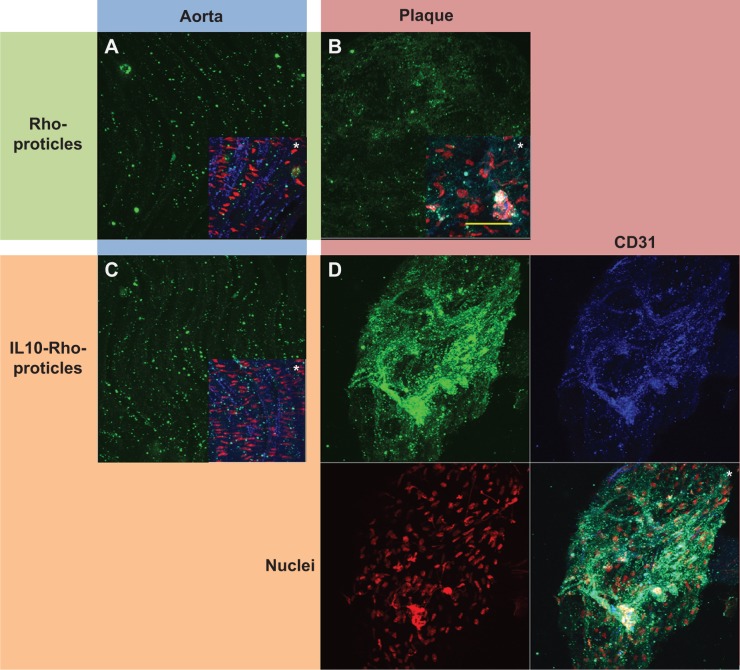
Figure 2.
The staining signal of IL10-coated protamine- and oligonucleotide-based NPs, detected on atherosclerotic-plaques and compared to controls.
Notes: Aortic sections of ApoE-deficient mice were incubated with rhodamine-labeled proticles for control (A) and (B), or coated with IL10 (C) and (D); NP signals are shown in green. Less-injured aortic areas are shown in (A) and (C) (blue area), atherosclerotic-plaques in (B) and (D) (red area). All images are depicted merged (*) with co-stainings for CD31 (endothelium) shown in blue and for the nuclei shown in red; images of the IL10-Rho-proticles stained plaque are also shown split. IL10-Rho-proticles were found to highly co-localized with the endothelium of atherosclerotic-plaques (bluish-green color). Bar indicates 50 μm.
Abbreviations: IL10, interleukin 10; NPs, nanoparticles; AS, atherosclerotic; ApoE, apolipoprotein E; Rho, rhodamine.

