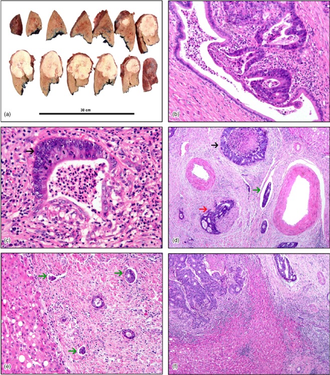
Figure 1.
(a) Gross specimen of hepatic resection showing a single tumour nodule. The distance between the front of the tumour and the margin of excision is marked with an arrowhead. Histopathology shows: (b) early invasion of the biliary epithelium by metastatic adenocarcinoma; (c) invasion of the biliary epithelium of a medium-sized duct by metastatic adenocarcinoma (arrowhead); (d) complete replacement of the biliary epithelium by adenocarcinoma (black arrowhead) with perineural invasion (red arrowhead) and adenocarcinoma cells within vascular spaces (green arrowhead); (e) multiple lymphatic spaces containing metastatic adenocarcinoma cells (green arrowheads), and (f) metastatic adenocarcinoma within the lumen of a blood vessel (top right). [Haematoxylin and eosin stain; original magnification (b) ×20, (c) ×20, (d) ×10, (e) ×10, (f) ×10]

