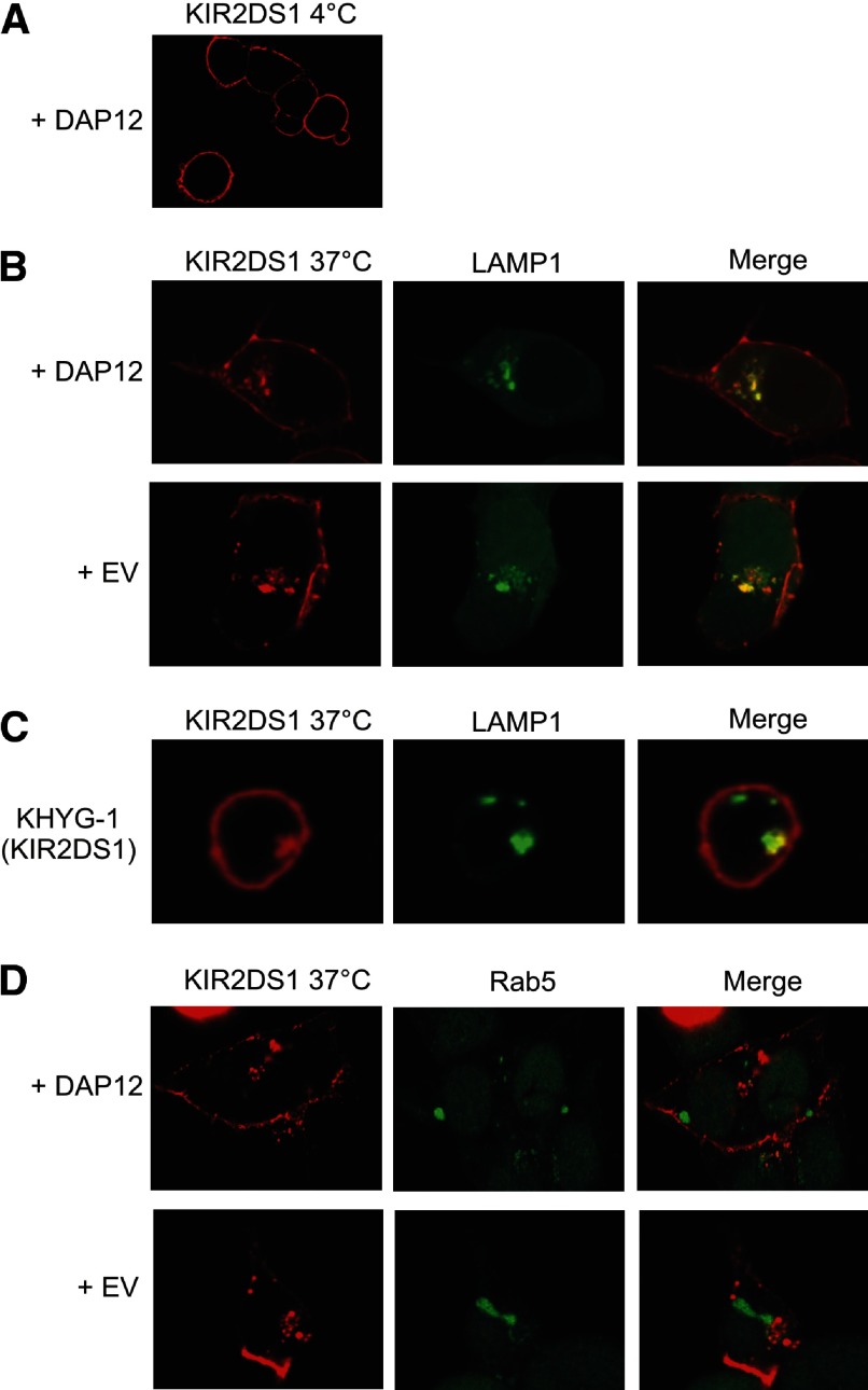
Figure 8. Confocal microscopy shows that internalized KIR2DS1 localizes to LAMP1-associated lysosomal compartments.
(A) KIR2DS1 was transfected with DAP12 into HEK293T cells and probed with a KIR2DS1-specific antibody at 4°C. (B) HEK293T cells were transfected with KIR2DS1 and empty vector or DAP12, along with a vector encoding LAMP1-GFP. The cells were probed with an APC-conjugated KIR2DS1 antibody for 2 h at 37°C, resulting in internalization of the receptor. The merged images to the right show the overlay of the KIR2DS1 and LAMP1 images. (C) KHYG-1 cells stably expressing KIR2DS1 were probed with a KIR2DS1-specific antibody at 37°C. The cells were then fixed, permeabilized, and stained using a LAMP1-specific antibody. (D) HEK293T cells were transfected with a vector encoding Rab5-GFP, along with KIR2DS1 with empty vector or DAP12. The merged images in the rightmost column display the overlay of the KIR2DS1 image with the LAMP1 or RAB5 images obtained. The image acquired after 4°C incubation was taken with a magnification of 100×, whereas the remaining 37°C images were obtained with a 200× magnification. The images represent results observed in 10 fields in each of three independent experiments.

