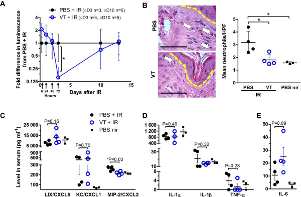
Figure 3.

Early local and systemic inflammatory signs after irradiation. (A) Irradiated skin MPO detection by bioluminescence in irradiated and PBS/VT-treated mice 6 h, 24 h, 48 h, 72 h, 10 days and 13 days after 35 Gy. Expressed as mean ± SD. (B) (Left) Representative H&E-stained skin sections of 40 Gy irradiated and PBS/VT-treated mice on day 5. Dashed line indicates epidermal and dermal boundary. Arrows point out neutrophils under 400x magnification (scale bars = 62.5 μm) and their counts are expressed as the mean of 20 HPFs per mouse ± SD (right). Serum cytokine levels in blood harvested 5 days after 40 Gy skin irradiation: (C) neutrophil-recruiting chemokines, (D) general pro-inflammatory driver cytokines and (E) the pro-inflammatory/neutrophil mobilizing cytokine IL-6, expressed as mean ± SEM. * signifies P < 0.05. Non-significant P-values are also included to aid in evaluation of differences between cytokine levels.
