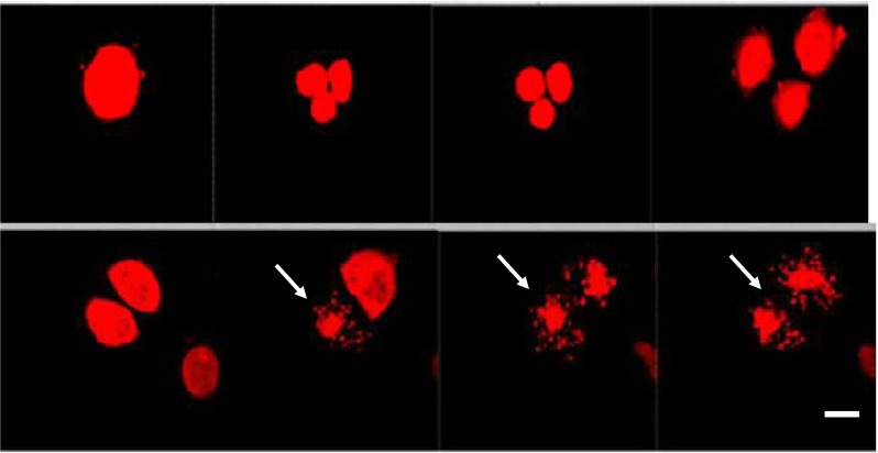
Fig. 6.

CTCs isolated from a tumor bearing mouse (orthotopically-transplanted human H460-RFP), cultured in vitro, imaged by time lapse imaging for 72 h. An irregular mitosis of one dividing cell (in the multinuclear stage) is shown on the picture. Subsequently three daughter cells developed. Each daughter-cell divided within the next mitotic cycle into 3 cells. Afterwards an apoptosis process started (see arrows). The CTCs were kept in RPMI medium with FBS. Scale bar 10 μm (Leica confocal microscope TCS SP5 AOBS)
