
Figure 3.
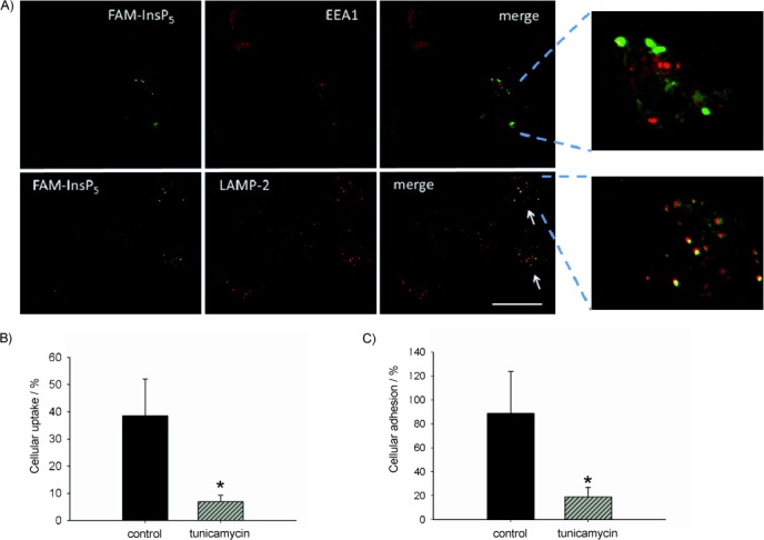
Uptake of FAM-InsP5 (5) into H1299 cells. A) H1299 cells were grown to 50 % confluence and treated with 5 (20 μm). After 16 h the cells were washed, fixed with paraformaldehyde and treated with primary antibodies against marker proteins for early endosomes (EEA1) and LAMP-2 (lysosomes), which were then detected with the aid of secondary antibodies labelled with Alexa Fluor 568. The yellow colour in the merge micrographs indicates colocalisation between 5 and the corresponding marker proteins (see arrows). Scale bar: 10 μm. B) Inhibition of FAM-InsP5 (5) uptake by tunicamycin. H1299 cells grown to 50 % confluence were either treated with tunicamycin (25 μm) for 6 h or remained untreated (control). After washing of the cells twice with PBS, they were treated with 5 (20 μm) and 16 h later they were washed and fixed with paraformaldehyde. Internalisation of 5 was analysed as described in Figure 1. C) Cells were treated as described in B. Then, the washed cells were treated with 5 (20 μm) for 50 min at 4 °C and the fluorescence at the cell surface was analysed in the XY-plane. Percentage cellular adhesion of 5 was calculated from ten micrographs, and the results are depicted in the histogram (mean values±SEM) * p<0.0001.