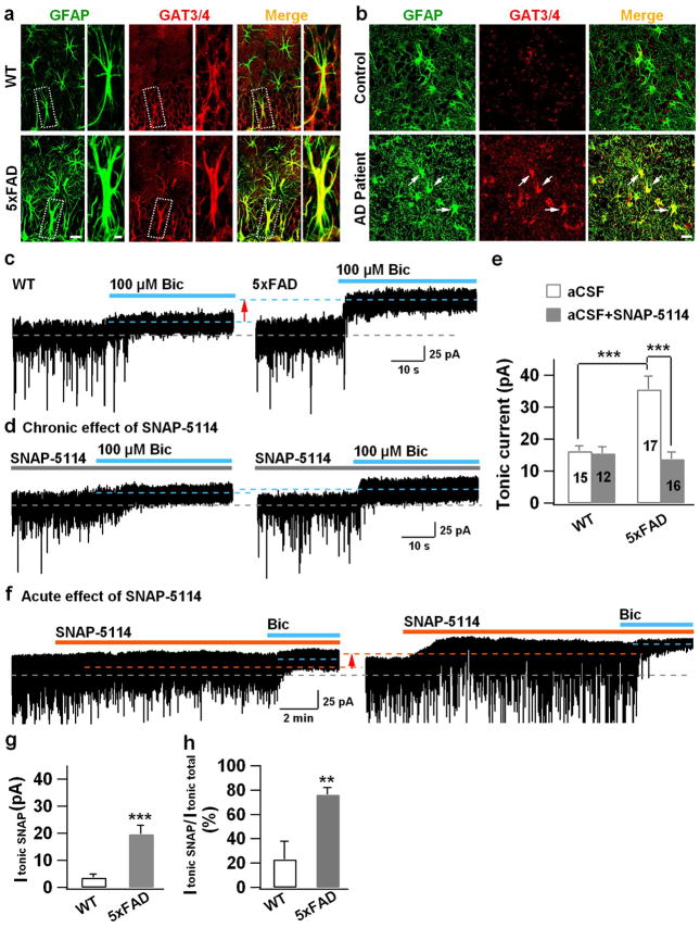
Figure 5. Increase of GAT3/4 in AD reactive astrocytes and contribution toward enhanced tonic GABA current in dentate granule cells.
(a) Significant increase of GAT3/4 immunoreactivity (red) in the dentate astrocytes (GFAP, green) in 5xFAD animals (bottom row), compared to controls (top row). n = 6 mice for WT or AD group. Scale bar = 20 μm (5μm in enlarged image). (b) Human dentate astrocytes (GFAP, green) in AD patients (bottom row) also showed remarkable increase of GAT3/4 staining (red, arrow). n = 6 for both control and AD groups. Scale bar = 20 μm. (c) Dentate granule cells in 5xFAD animals (right panel) showed enhanced tonic current, revealed by GABAA-R antagonist bicuculline (Bic), compared to controls (left panel). (d) Chronic blockade of GAT3/4 with specific inhibitor SNAP-5114 (50 μM, 2 hr) significantly diminished the tonic current in dentate granule cells in 5xFAD animals (right panel), compared to that in WT mice (left panel). Synaptic currents were truncated for better view of the small tonic current. (e) Quantified data of panel c–d (ANOVA with Tukey post hoc tests) and cell number is shown in histograms. (f) Acute application of SNAP-5114 also blocked the majority of the tonic current in dentate granule cells in 5xFAD animals, but showed little effect in WT dentate granule cells. (g, h) Quantified data in panel f, showing a major contribution of GAT3/4 toward the tonic current in 5xFAD dentate granule cells (Student’s t test). n = 12 for WT and 13 for 5xFAD group. Data are presented as mean ± s.e.m., ** P < 0.01, *** P < 0.001.

