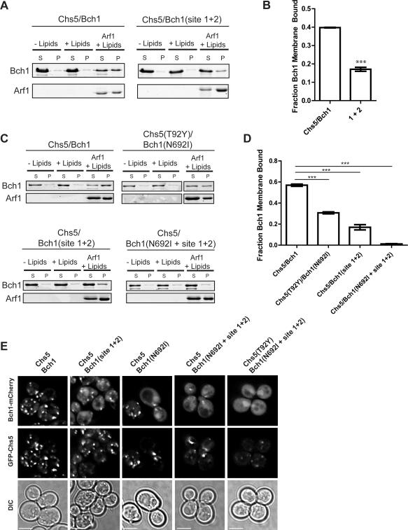
Figure 4. Exomer membrane recruitment occurs through multiple interactions with Arf1 and membranes.
A) Liposome pelleting assay to test Arf1-dependent membrane binding of Chs5(1-299)/Bch1 mutants.
B) Quantification of panel (G), n = 3, with significance determined by an unpaired t-test.
C) Liposome pelleting assay to determine the contributions of multiple exomer interfaces. Chs5-T92Y and Bch1-N692I are Arf1 interface mutations, site 1+2 is the membrane interface mutation.
D) Quantification of the data shown in panel (C), n = 3.
E) Plasmids expressing GFP-Chs5 and Bch1-mCherry constructs were introduced into a chs5Δchs6Δbud7Δbch1Δ strain and imaged. Scale bar, 2 μm.

