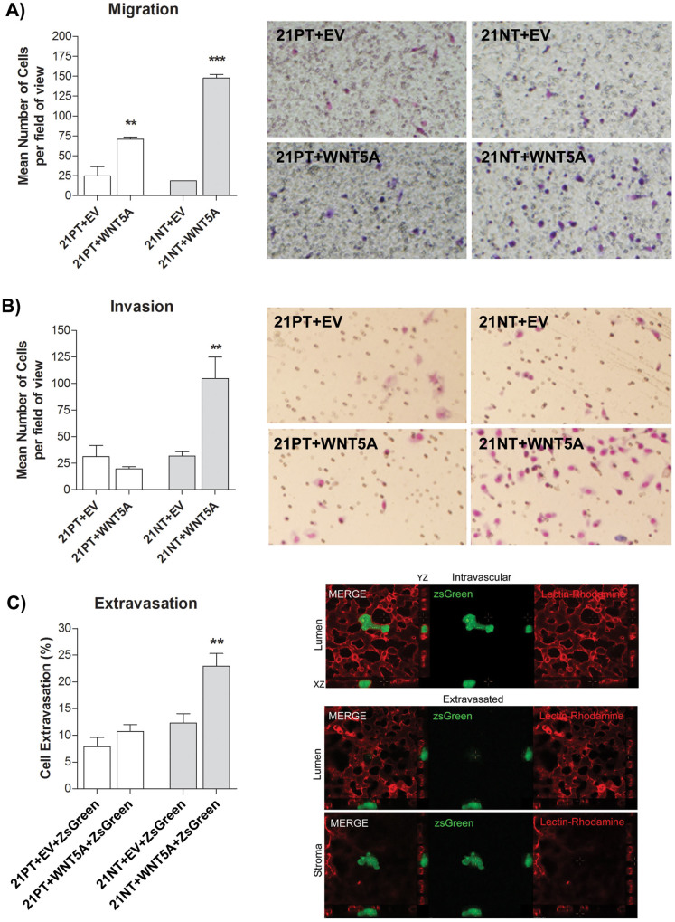
Figure 3. WNT5A promotes migration of 21PT and 21NT, but increases invasion and extravasation of 21NT cells only.
(A) Transwell migration assay (transwells coated with gelatin) using 21T transfectants towards 10% FBS for 5 hours. Both 21PT + WNT5A cells and 21NT + WNT5A cells showed an increase in the number of cells that migrated to the underside of transwell membranes, compared to their respective empty vector control. Representative transwell images are shown (right panel). (B) Transwell invasion assay (transwells coated with Matrigel) using 21T transfectants towards 10% FBS for 20 hours. 21NT + WNT5A cells showed an increase in the number of cells that invaded to the underside of the transwell membrane compared to the empty vector control. However, WNT5A overexpression did not increase invasion of 21PT cells. Representative transwell images are shown (right panel). (C) In vivo (chick embryo) cancer cell extravasation assay using 21T transfectants labeled by transduction with ZsGreen. WNT5A overexpression significantly increased the ability of 21NT, but not 21PT cells to extravasate. Representative (21NT transfected) cells are shown in the intravascular (luminal) and extravascular (stromal) state (right panel). To analyze groups a one-way ANOVA was performed with Tukey's post hoc test and P < 0.05 was considered statistically significant. **P < 0.01; ***P < 0.001.

