
Fig. 7.
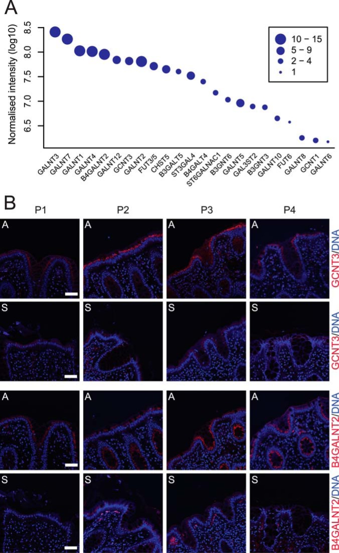
Relative abundance and immunohistochemistry of the identified glycotransferases involved in O-glycosylation. A, relative abundance of the identified glycosyltransferases combined for all colon segments showing a 100-fold difference between the most and least abundant transferase. The size of the points indicates the number of uniquely identified peptides assigned to each glycosyltransferase. B, immunostaining (red) of the glycosyltransferases B4GALNT2 and GCNT3 in tissue sections from ascendant (a) and sigmoid colon (s) of the four patients included in this study (P1–P4). Nuclei were stained with Hoechst (blue). Bar = 50 μm.