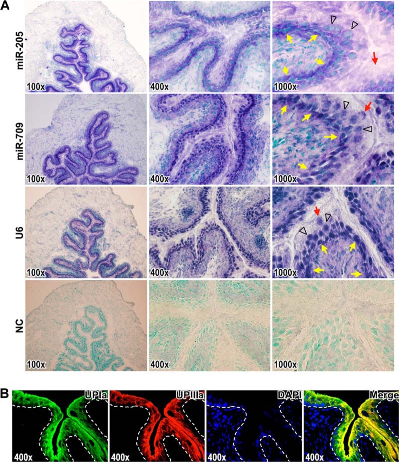
Fig. 2.

miR-205 is expressed in the lower undifferentiated layers of urothelium. A, In situ hybridization detection of miR-205 and miR-709 on mouse bladder sections. Images represent sections hybridized with antisense probes corresponding to miR-205, miR-709, positive control U6, and negative control (NC) in magnifications 100 x, 400 x, and 1000 x, respectively. The hybridization signals are in purple blue. Nuclei were counterstained with methyl green dye. The yellow arrows indicate the epithelium-mesenchyme boundary. The black arrowheads indicate the intermediate cells. The red arrows indicate the superficial umbrella cells. B, The urothelial differentiation was determined with the specific differentiation markers, uroplakin Ia (UPIa), and uroplakin IIIa (UPIIIa), in upper urothelial cell layers. Localization of UPIa (green) and UPIIIa (red) were observed by immunofluorescence (yellow in merged image). The dashed lines indicate the epithelium-mesenchyme boundary. DAPI-stained nuclei are shown in blue.
