
Fig. 1. Surround inhibition during motor imagery.
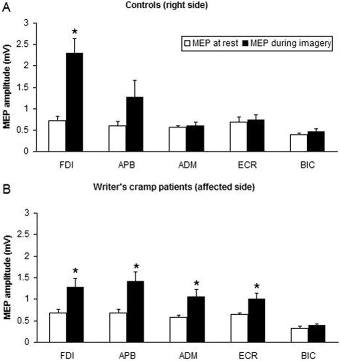
Changes in mean motor evoked potential (MEP) size during motor imagery on the right side of healthy controls and on the affected side of patients. TMS was always given to the motor cortex contralateral to the imagined task. MEP size during motor imagery (MI) compared with rest condition recorded from different target muscles of the right upper limb after stimulation of the left hemisphere in controls (A) and patients with writer’s cramp (B). The bar chart illustrates the mean peak-to-peak amplitude (mV) of MEPs recorded at rest (open columns) and during MI (black columns). Each error bar equals standard error of the mean (SEM). MI elicited an attenuated and less focal increase in MEP amplitude in patients than in controls. FDI, first dorsal interosseus; APB, abductor pollicis brevis; ADM, abductor digiti minimi; ECR, extensor carpi radialis; BIC, biceps. *P < 0.05. (from: Quartarone A, Bagnato S, Rizzo V, Morgante F, Sant’Angelo A, Crupi D, Romano M, Berardelli A, Girlanda P. Corticospinal excitability during motor imagery of a simple tonic finger movement in patients with writer’s cramp. Mov Disord. 2005 Nov;20(11):1488–95).