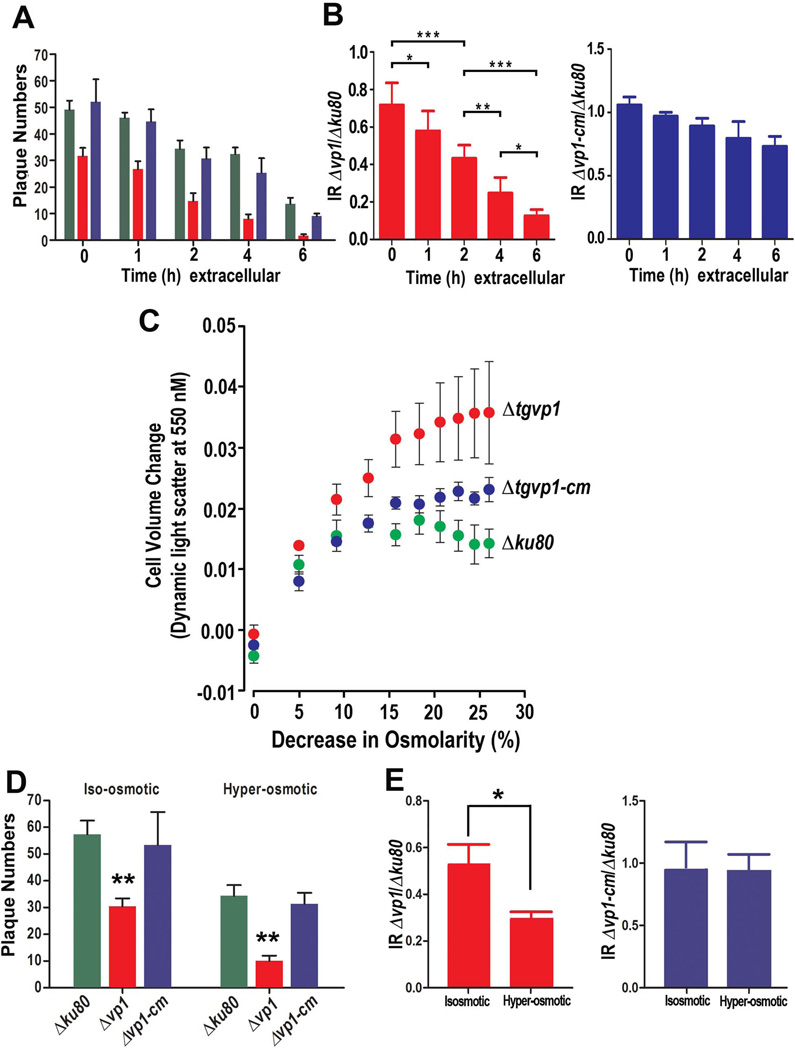
Fig. 6.
Extracellular Δvp1 parasites are more sensitive to environmental stress. (A) Plaque numbers after pre-incubation of parasites in Ringer buffer for different lengths of time (0, 1, 2, 4 or 6 h). (B) Ratio (Invasion Ratio, IR) between the number of plaques formed by Δvp1 and Δku80 after pre-incubation in an extracellular buffer for 0, 1, 2, 4 and 6 h (red columns). Blue columns show the same ratio calculation for Δvp1-cm and Δku80. All strains show decreased number of plaques after long extracellular incubation but the decrease in the number of plaques produced by tachyzoites from Δvp1 clone was more dramatic than that produced by tachyzoites of Δku80 and Δvp1-cm clones. (C) Effect of hyposmotic conditions on volume changes of tachyzoites from different clones as determined by light scattering. Data are the combined results of three independent experiments. (D) Plaque numbers after pre-incubation of parasites under isosmotic or hyperosmotic conditions. (E) Ratio (IR) of the number of plaques formed by Δvp1 and Δku80 submitted to hyperosmotic stress compared to the same ratio for parasites incubated for the same length of time in an isosmotic buffer (red columns). The blue columns show the same calculation between the number of plaques formed by the Δvp1-cm and Δku80 tachyzoites. Tachyzoites from Δvp1 clone were less resistant to this treatment than tachyzoites from Δku80 and Δvp1-cm clones and this is evident in the lower ratio obtained under hyposmotic conditions. Data from (A,B) and (D,E) were analyzed by GraphPad Prism 5. To compare two groups of data we used One-Way ANOVA test. *, p <0.05; **, p <0.01; ***, p <0.001.

