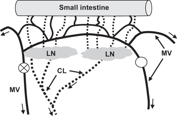
Fig. 1.

Experimental induction of mesenteric venous hypertension. The veins (solid lines) draining the small intestine are arranged in arcades so that complete obstruction (○) of one mesenteric vein (MV) and constriction (represented as circle with X) of an adjacent MV does not stop blood flow, but rather redirects flow and increases local venous pressure. The resulting increase in intestine microvascular pressure causes microvascular filtration and interstitial water volume to increase. As a result, lymph flow from the intestine and through the lymph nodes (LN) and postnodal collecting lymphatic vessels (CL) increases. Arrows indicate direction of flow for venous blood and lymph.
