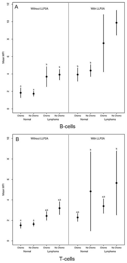
Fig. 3.
Mean ± 2 SE median fluorescence intensity (MFI) in B-cells (A) and T-cells (B) with respect to LLP2A labeling, lymphoma status (normal or lymphoma) and whether the dog received chemotherapy (chemo, no chemo). Groups with the same letter do not differ significantly (p > 0.05). B-cell lymphoma showed higher affinity to LLP2A than non-neoplastic lymphocytes. Both of these groups had higher MFI than unlabeled lymphoma cells. Chemotherapy significantly decreased the mean MFI in B-cell lymphoma. There was increased affinity of LLP2A to T-cell lymphoma although this was not significant.

