
Fig 5. Dendritic spine pruning defects and ASD like behaviors in Atg7CKO mice.
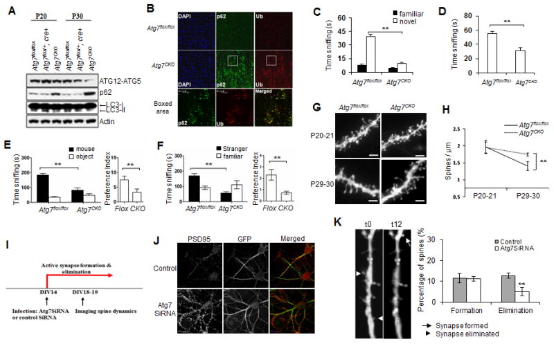
(A)Western blot analysis of autophagy markers in the Atg7CKO cortex. Brain homogenates from P19-20 and P29-30 mice were immunoblotted with antibodies against Atg12-Atg5, LC3 and autophagy substrate p62. Data shown are representative of three separate experiments. Loss of autophagy was indicated by a decrease in levels of Atg5-12 conjugation and LC3-II protein, and an increase in p62 protein; (B) Immunofluorescent labeling of p62 and ubiquitin (Ub) in P30 Atg7CKO mouse cortex. Scale bar: 10 μm. (C-F) ASD like social behaviors in Atg7CKO mice. Atg7flox/flox males, n=15; Atg7CKO males, n=13; (C) Novel object recognition test showing time spent sniffing a familiar object vs. a novel object; (D) Dyadic social interaction test showing the time testing mice spent sniffing a stimulus mouse. ** Compared to Atg7flox/flox; p<0.01; unpaired t-test; (E) Sociability in the 3-chamber test showing time spent (left) and preference (right) for a stimulus mouse or an object; (F) Social novelty in the 3-chamber test showing time spent (left) and preference (right) for sniffing a stranger mouse vs. a familiar mouse. Compared to wt, ** p<0.01 (unpaired t test); (G) Dendritic segments from Atg7flox/flox and Atg7CKO pyramidal neurons at P19-20 and P29-30. n=7-10 animals per group. Scale bar: 2 μm; (H) Fewer spines were pruned in Atg7CKO mice. ** Compared to P29-30 Atg7flox/flox, p<0.01 (2-way ANOVA, Bonferroni post hoc test). (I) Timeline of infection and spine analysis; (J) Cultured control and Atg7 SiRNA lentiviral infected mouse hippocampal neurons at DIV20, visualized by GFP and PSD95 fluorescence. Atg7SiRNA expressing neurons exhibited more PSD95 puncta than controls; (K) Spine formation and elimination in control and Atg7SiRNA infected neurons during a 12-hour time window at DIV19-20.