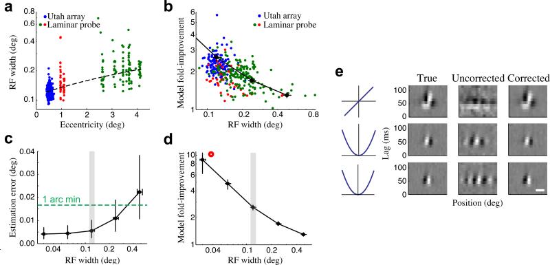
Figure 6. Dependence of eye position and model estimation on RF size.
(a) Recordings performed across different eccentricities showed a large range of RF widths at a given eccentricity, as well as the expected increase in average RF width with eccentricity. Each point represents the RF width and eccentricity of a given single- or multi-unit (N = 76/274 SU/MU), color-coded by electrode array and recording location (blue: Utah array, fovea, N = 132); red: linear array, fovea, N = 48; green: linear array, parafovea, N = 170). The dashed line shows the linear fit between eccentricity and RF size (note semi-log axes). (b) Cross-validated improvements in model performance were larger for neurons with smaller RFs (correlation = −0.53, p = 0). Units color-coded as in (a). A similar relationship was also found using simulated neural populations (see below: black trace). (c) Simulations, where ‘ground truth’ eye positions are known, were performed for V1 populations (N = 101 units) having a range of RF widths. The accuracy of inferred eye positions increased for populations with smaller RFs, and remained below 1 arc minute even for populations with substantially larger RFs. Error bars show the median and inter-quartile range, and the RF widths of foveal neurons recorded on the planar array are indicated by the gray region. (d) For the same simulated V1 populations, improvements in model fits became more dramatic for neurons with smaller RFs, although they remained substantial even for neurons with much larger RFs. Note that this is the same trace as in (b). (e) Stimulus filters can be recovered even for neurons with the smallest RF widths (left), as demonstrated for a simulated neuron with an RF width of 0.037 deg (red circle in (d)). Here, fixational eye movements almost entirely obscure the model estimates without eye position correction (i.e., assuming perfect fixation) (middle), but using the inferred eye positions recovers the structure of the neuron's stimulus filters (right). Horizontal scale bar is 0.05 deg.

