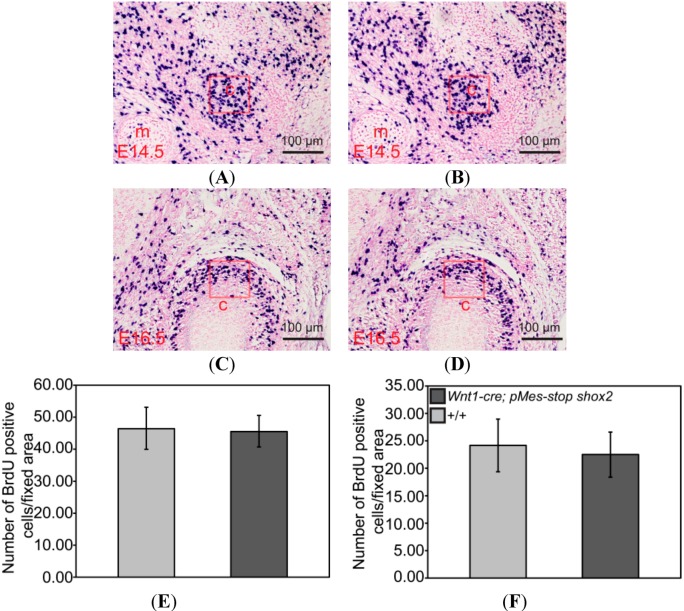
Figure 6.
Changes of proliferation in the TMJ of Wnt1-Cre; pMes-stop Shox2 mice. (A–D) BrdU labeling of cell proliferation in the condylar condensation of an E14.5 (A,B) and E16.5 (C,D) in wild type and Wnt1-Cre; pMes-stop Shox2 embryo; (E,F) Comparison of numbers of BrdUpositive cells in the fixed area (red frame) of the condylar primoridain between wild type (E) and Wnt1-Cre; pMes-stop Shox2 embryos (F). Standard deviation is shown as error bars.c, condyle; and m, Meckel’s cartilage.

