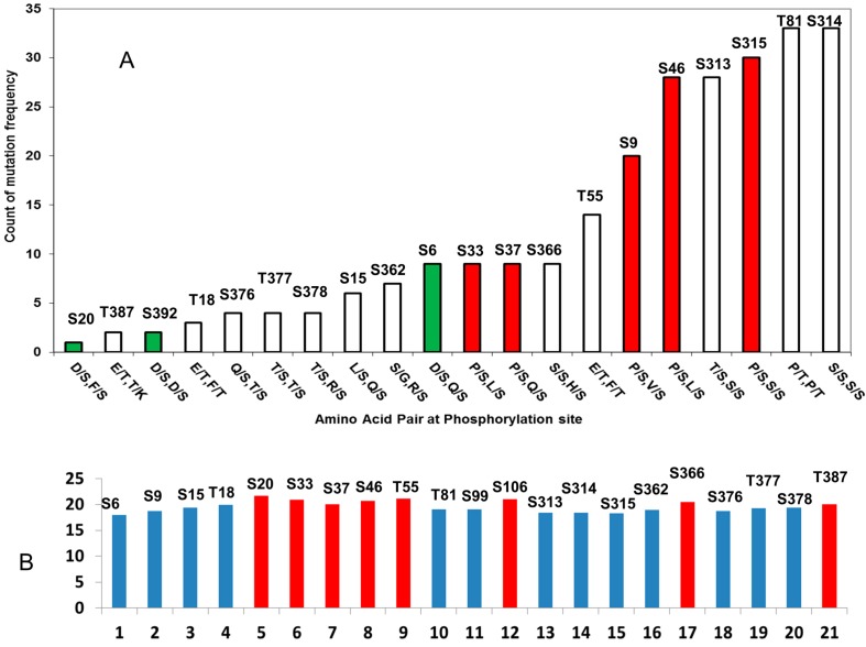
Figure 4.
p53 phosphorylation motifs can be characterized by amino acids adjacent to the phosphorylation motif and the propensity of structural disorder of the seven residue phosphorylation motif. (A) Phosphorylation motifs with D/S pattern (green bar) have fewer mutations than the motif with the P/S pair (red bar); (B) Phosphorylation motifs are structurally disordered (blue bar), some motifs, which are less disordered (red bar), have more secondary structure characteristics.

