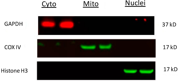
Figure 7.
Western blots showing purity of subcellular fractions. Cytosolic, mitochondrial and nuclear fractions were isolated according to the protocol described in the Experimental section and equal amount of proteins (20 µg) were loaded side by side on the same gel. The membrane was probed separately with three primary antibodies against GAPDH, COX IV and Histone H3. A single band for GAPDH at approximately 37 kD in the cytosolic fraction, an obvious band for COX IV at approximately 17 kD in the mitochondrial fraction, and a band for Histone H3 closing to 17 kD in the nuclear fraction was observed. There was no obvious contamination among different fractions.

