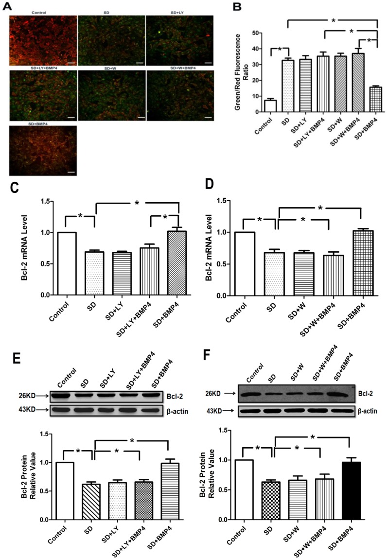
Figure 4.
BMP4 relieved mitochondrial potential reduction and induced Bcl-2 expression in PASMCs through the PI3K/AKT pathway. (A) The cells were stained with JC-1 probe and imaged by a fluorescentmicroscope. The individual red and green average fluorescence intensities are expressed as the ratio of green to red flintensitie. The increase of fluorescence ratio, which is represented in the bars, correlates with an increase in mitochondrial depolarization. Representative photographs of JC-1 staining in different groups. Scale bar = 10 µm; (B) Quantitative analysis of the shift of mitochondrial green fluorescence to red fluorescence among groups; (C,D) Quantitative real-time PCR was performed to quantify Bcl-2 expression. BMP4 increased the Bcl-2 mRNA expression induced by serum deprivation through PI3K/AKT pathway. Data were collected from three independent experiments with three to four samples in each; (E,F) The expression of Bcl-2 increased by BMP4 was partly inhibited by LY294002 and wortmannin (PI3K/AKT inhibitors). All values are denoted as means ± SEM from three or more independent batches of cells. “SD” means serum deprivation; “LY” means LY294002; “W” means wortmannin. * p < 0.05.

