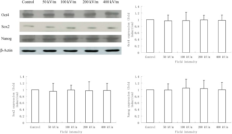
Figure 3.
Western blotting analysis of Oct4, Sox2 and Nanog expression in HUES-17 human embryonic stem cells (hESCs) after exposed to PEMF at different electric field intensities (control group, 50, 100, 200, 400 kV/m). β-Actin was used as an internal control. Protein level of each gene was quantitated by densitometry and plotted as fold induction. Results were obtained from three independent experiments and expressed as mean ± SD (n = 3 in each experiment).

