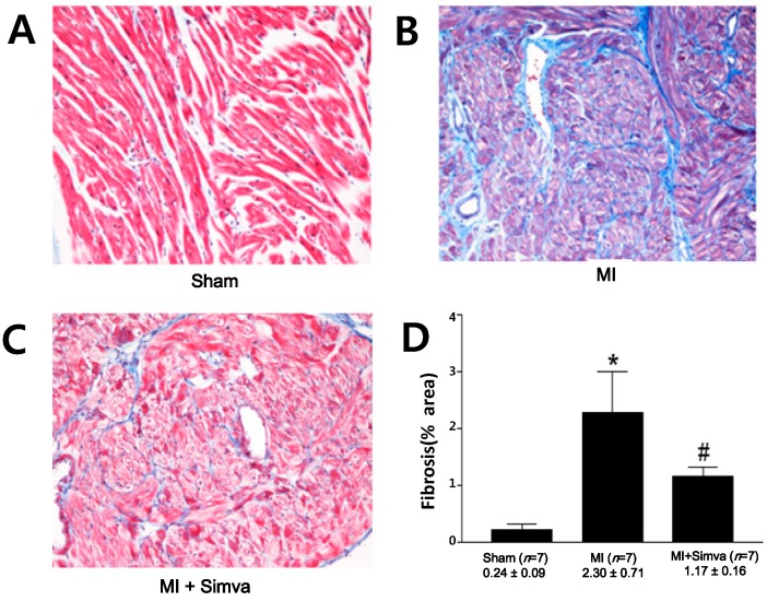
Figure 4.
Masson’s trichrome staining of the left atrium. The amount of fibrosis was lower in the sham (A) than the MI group (B); but this increase was attenuated in the MI + simvastatin (C) group. Blue represents fibrosis. (D) The effect of simvastatin on fibrosis of the left atrium. * p = 0.001 sham vs. MI and MI + simvastatin, # p = 0.001 MI vs. MI + simvastatin.

