Figure 4.
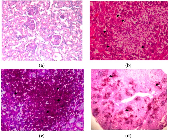
Kidney tissues on 72 h post-infection with C. albicans, (a) uninfected kidney; Haematoxylin-eosin (H&E) stain; original magnification, ×400 (Tissues were normal, no congestion of tubules, inflammation, haemorrhage and glomeruli); (b) H&E stain; original magnification, ×400 (Arrows indicated a large polymorphonuclear infiltrate is demonstrated in the abscess in the center of the photomicrograph); (c) Periodic acid Schiff (PAS) stain; original magnification, ×400 (Arrows indicated hyphae and fungal bodies of C. albicans); and (d) PAS stain; original magnification, ×40 (Arrows indicate that here there is confluent invasion of the hyphae and fungal bodies in the kidney).

